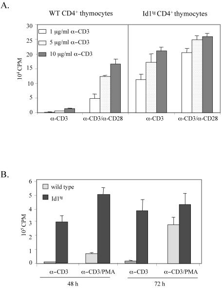
FIG. 4.
Costimulation-independent proliferation of CD4+ thymocytes from Id1 transgenic mice. (A) Sorted CD4+ thymocytes were plated in triplicates (2 × 10⁵ cells per well) in a 96-well plate coated with indicated concentrations of anti-CD3ɛ antibody with or without soluble anti-CD28 MAb (2 μg/ml) and incubated for 48 h. Cells were pulsed with 1 μCi of [3H]thymidine per well for the last 18 h of incubation. The amount of [3H]thymidine incorporated by proliferating thymocytes was measured by scintillation counting. (B) Sorted CD4+ thymocytes from wild-type and Id1 transgenic mice were stimulated with plate-bound anti-CD3ɛ antibody (10 μg/ml) in a 96-well plate with or without PMA (2.5 ng/ml) for 48 or 72 h. [3H]thymidine incorporation was measured as described for panel A.

