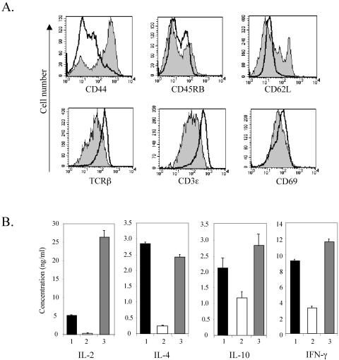
FIG. 5.
CD4+ thymocytes from Id1 transgenic mice do not display the phenotype of memory T cells. (A) Surface marker expression in CD4+ thymocytes. Thymocytes from wild-type and Id1 transgenic mice were stained with anti-CD4 and anti-CD8 plus one of the antibodies specific for the indicated antigens. The levels of the indicated surface antigens on gated CD4 SP thymocytes are shown in histograms. Solid lines represent wild-type thymocytes, and shaded areas designate Id1 transgenic thymocytes. (B) Cytokine secretion by activated CD4+ thymocytes. CD4+ thymocytes (2 × 10⁵ cells per well) were stimulated for 48 h with plate-bound anti-CD3ɛ (10 μg/ml) with or without soluble anti-CD28 monoclonal antibody (2 μg/ml). Culture media were collected and analyzed for the indicated cytokines by enzyme-linked immunosorbent assay. Bars: 1, wild-type CD4+ thymocytes stimulated with anti-CD3ɛ and anti-CD28; 2, Id1 transgenic CD4+ thymocytes stimulated with anti-CD3ɛ alone; 3, Id1 transgenic CD4+ thymocytes stimulated with anti-CD3ɛ and anti-CD28.

