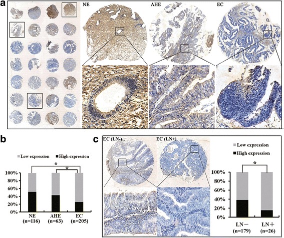
Fig. 1.

Expression of MIIP is reduced in human EC specimens. a MIIP expression was evaluated by immunohistochemical staining on TMAs. The respective images in the same TMAs showed that MIIP expression was lower in EC than those in NE and AHE. b Statistical analysis revealed that MIIP expression was highest in NE, lower in AHE, and lowest in EC. c Loss expression of MIIP was related to lymph node metastasis in EC. Left panel: Shown are representative images of MIIP expression in EC tissues with or without lymph node metastasis. Right panel: Statistical analysis revealed that low MIIP expression was correlated with lymph node metastasis in EC patients. Asterisk indicates P < 0.05. See also Table 1
