Fig. 2.

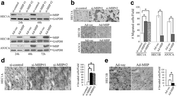
MIIP inhibits EC cell migration and invasion. a Western blot shows that MIIP was knocked down by two different siRNAs against MIIP when compared to control at 24 and 48 h. And MIIP expression was forced in HEC1B and AN3CA cells by infection with an adenovirus containing MIIP (Ad-MIIP) or control adenovirus (Ad-Ev) at 24, 48, or 72 h. b, c Modulation of EC cell migration by MIIP in a transwell migration chamber. b Representative photographs revealed knockdown of MIIP enhanced HEC1A cell migration and overexpression of MIIP inhibited HEC1B and AN3CA cell migration (magnification ×200). c Data are expressed as means ± SD of cells per 10 high-power fields from three separate experiments. d, e Modulation of EC cell invasion by MIIP in a transwell invasion chamber. d Left: Representative images of cells on the filter surface of HEC1A (×200 magnification). Right: Quantitative measurement of invaded HEC1A cells. Data are represented by the mean ± SD of cells per 10 high-power fields from three separate experiments. e Left: Representative images of cells on the filter surface of HEC1B (×200 magnification). Right: Quantitative measurement of invaded HEC1B cells. Asterisk indicates P < 0.01
