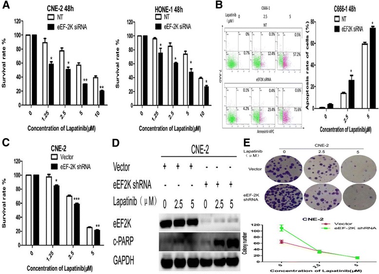
Fig. 3.

Silencing of eEF-2 kinase expression by RNA interference augments lapatinib-induced apoptosis in NPC cells. a and b NPC cells were transfected with a non-targeting RNA (NT) or siRNA targeting eEF-2 kinase (eEF-2 K siRNA) followed by treatment with lapatinib or DMSO for 48 h. a Cell viability was assessed by the CCK-8 assay. b Annexin V-APC/7-AAD double staining was performed to detect apoptotic activity. Results are displayed as histograms. Each bar represents the mean ± standard deviation. *, P<0.05 and **, P<0.01. c, d and e NPC cells were transfected with an empty vector control (Vector) or a shRNA targeting eEF-2 kinase (eEF-2 K shRNA) followed by treatment with lapatinib or DMSO control for 48 h. c Cell viability was assessed by the CCK-8 assay. d Cleaved PARP and eEF-2 kinase levels were examined by Western blot analysis. GAPDH was used as a loading control. Results are displayed as histograms. Each bar represents the mean ± standard deviation. *, P<0.05;**, P<0.01 and ***, P<0.001. e Colony formation was measured. One representative experiment is shown (CNE-2 cells). Results are displayed as line charts to compare the decreasing trends in colony number
