Fig. 5.

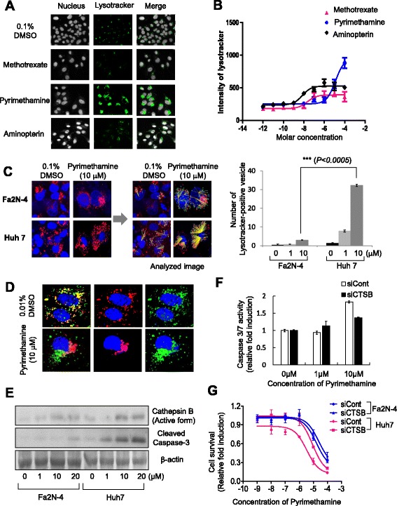
Pyrimethamine induces lysosomal modification and release of cathepsin B in HCC cells. a Fluorescence images of lysosomes (LysoTracker® Green DND-26; green) and b intensity of LysoTracker in the Huh7 cell line after anti-folate drug treatment (x-axis indicates molar concentration). c Lysosome staining images (LysoTracker® Red DND-99, red) of the Fa2N-4 and Huh7 cell lines after pyrimethamine treatment (left; 48 h, 10 μM) and image analysis of LysoTracker Red-positive vesicle number (right). Graph represents LysoTracker-positive vesicles in the Fa2N-4 and Huh7 cell lines after pyrimethamine treatment (bottom). d Translocation of cathepsin B was detected in the Huh7 cell line after pyrimethamine treatment (48 h, 10 μM) and e expression of active cathepsin B and active caspase-3 detected by Western blot analysis after incubation with pyrimethamine at the indicated concentration. f The level of active caspase 3/7 in Huh7-siCont and Huh7-siCTSB cells after pyrimethamine treatment at 1, 10 μM concentration. g Drug response curves of Huh7-siCont, Huh7-siCTSB, Fa2N-4-siCont, and Fa2N-4-siCTSB cells after treatment of pyrimethamine at ten point concentrations (from 1 pM to 100 μM). All images and analyses were examined using the High Content Screening System. Experiments were performed in triplicate. Error bars indicate standard deviation. Images in the same panel were obtained with same magnification
