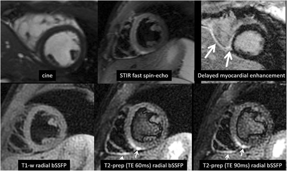
Fig. 4.

Patient presenting with chest pain and CMR findings consistent with myocarditis. Top row (from left to right): cine, T2-weighted STIR fast spin-echo, myocardial delayed enhancement using inversion recovery spoiled gradient-echo. Bottom row (from left to right): T1-weighted radial bSSFP, T2-weighted radial bSSFP with T2prep time = 60 ms, T2-weighted radial bSSFP with T2prep time = 90 ms. The T2-signal abnormality involving the inferior walls of the left and right ventricles is seen in the T2-weighted STIR fast spin-echo image, but is better delineated with T2-weighted radial bSSFP. Right ventricular trabeculations are also better delineated with the radial acquisitions. In this example, the blood pool was effectively nulled in the T2-weighted STIR fast spin-echo and T1-weighted radial bSSFP images, whereas the left ventricular blood pool has intermediate signal intensity in the T2-weighted radial bSSFP image due to suboptimal setting of the time delay following the dual-inversion preparation
