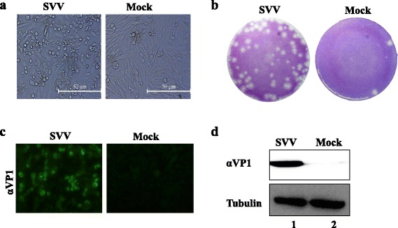
Fig. 2.

Identification of SVV HB-CH-2016 strain. a The cytopathic effect of BHK-21 cells infected with SVV HB-CH-2016 strain at 18 h post-infection. b Plaque morphology in BHK-21 cells infected with fourth-passage SVV HB-CH-2016 strain at 48 h post-infection. c Immunofluorescence assay (IFA) of BHK-21 cells infected with SVV HB-CH-2016 strain at 12 h post-infection. Cells were stained with primary antibody using home-made mouse anti-SVV VP1 polyclonal antibodies. d Western blot analysis of BHK-21 cells infected with SVV HB-CH-2016 strain at 12 h post-infection. Cells were stained with primary antibody using a home-made mouse polyclonal anti-SVV VP1 antibody and mouse anti-tubulin antibody
