Fig. 1.

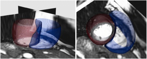
Two views of a short-axis CMR with the subject-specific 3D bi-ventricle model overlaid. The LV endocardium is shown in white, the LV epicardium is shown in red, and the RV endocardium in blue

Two views of a short-axis CMR with the subject-specific 3D bi-ventricle model overlaid. The LV endocardium is shown in white, the LV epicardium is shown in red, and the RV endocardium in blue