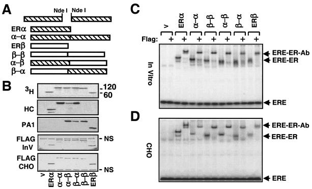
FIG. 3.
Construction and synthesis of fusion ERs. (A) Schematics of ER fusion receptor cDNAs. A PCR-generated NdeI restriction enzyme site at the 5′ or 3′ end of an ER was used to genetically fuse two ER cDNAs in tandem. (B) Synthesis of fusion ERs in vitro with the parent vector bearing no cDNA (V) or a cDNA for an ER was accomplished as described for Fig. 1. Equal amounts of in vitro reaction mixtures (5 μl) or of (10 μg of total protein) of cell extracts from transfected CHO cells (Flag-CHO) were subjected to SDS-7.5% PAGE. The in vitro samples were visualized by fluorography (3H). The same samples were also probed with the HC, the PA1, or the Flag antibody (Flag-InV). (C) ERE binding of in vitro or in situ (CHO)-synthesized fusion receptors. Equal amounts (2 μl) of reaction mixtures, with the exception of the mixture containing ERβ (10 μl), were incubated with radiolabeled ERE in the absence or presence of the Flag antibody. Reaction mixtures were electrophoresed by 5% nondenaturing PAGE. (D) Equal amounts of CHO cell extracts (10 μg) were subjected to EMSA. The results from a representative experiment of two independent experiments are shown.

