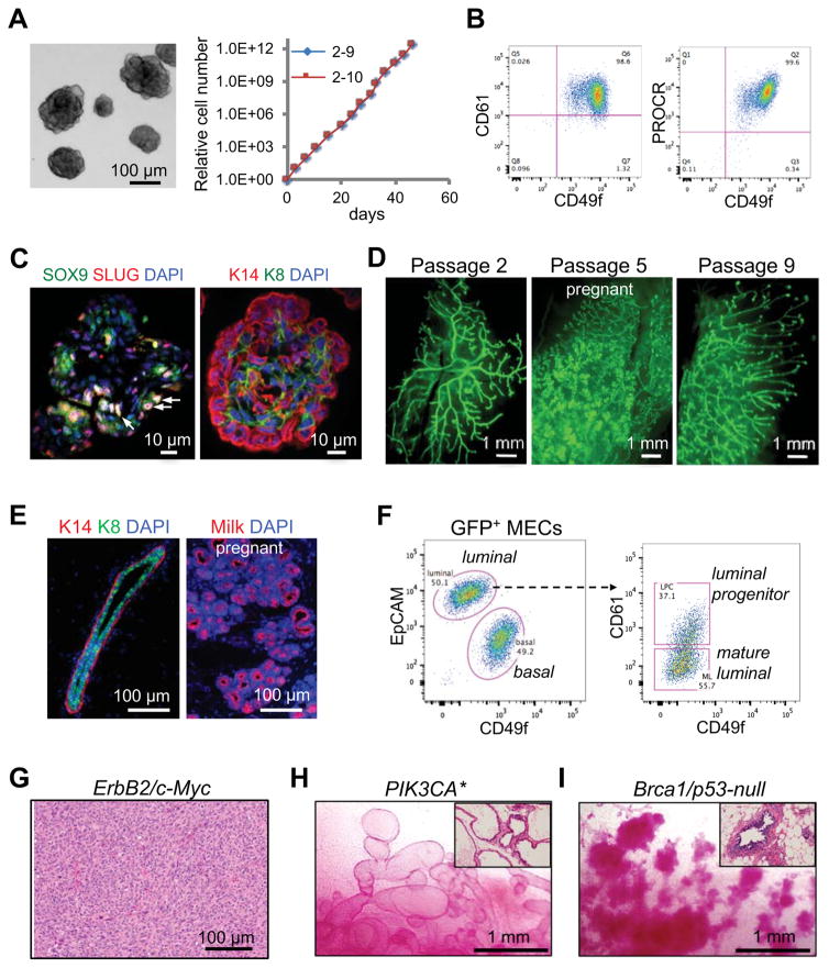
Figure 1. Rapid generation of somatic GEMMs for breast cancer by ex vivo expansion and modification of MaSCs.
(A) Long-term expansion of MaSC organoids. Left: a representative image of organoids. Right: Growth curves of two single cell-derived organoid clones.
(B) CD49f, CD61 and PROCR flow cytometric profiles of organoids.
(C) SLUG, SOX9 and cytokeratin immunostaining in organoids. The arrows indicate examples of SLUG+SOX9+ cells.
(D) Whole-mount images of mammary ductal trees regenerated by single cell-derived GFP+ organoids at the indicated passages.
(E) Immunofluorescence images of mammary ducts (left, virgin) and alveoli (right, pregnant) regenerated by organoids (passage 2).
(F) Flow cytometric profiles of mammary ductal trees regenerated by organoids (passage 8).
(G) A representative H&E image of poorly differentiated adenocarcinoma developed in Erbb2/MYC MaSC-GEMMs (with passage 3 organoids). Organoids were transplanted into NOD-SCID mice, and the mice were treated with doxycycline for 4 months.
(H) Representative images of carmine-stained mammary fat pads reconstituted by Rosa-CreERT2; Pik3ca* organoids (passage 3–5). Mice were treated with tamoxifen 6 weeks after transplantation and then analyzed 6 weeks later. The inset shows H&E staining of the outgrowths.
(I) Representative images of carmine-stained mammary fat pads reconstituted by Blg-Cre; Brca1floxed/floxed; p53−/− organoids (passage 5). The inset shows H&E staining of the outgrowths.
See also Figure S1.

