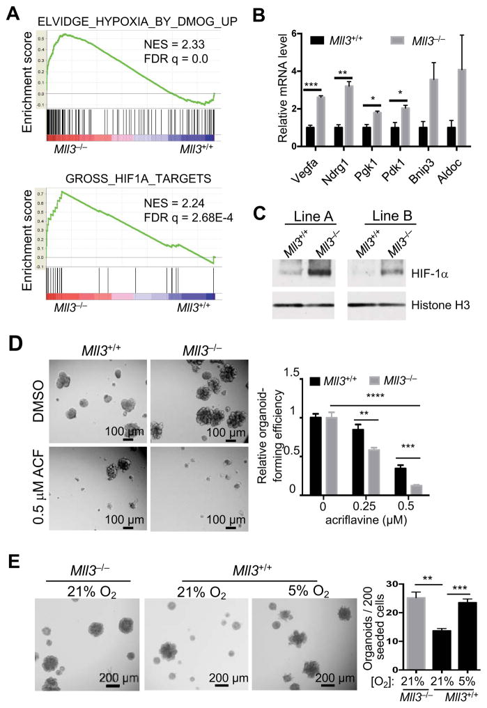
Figure 5. Activation of the HIF pathway mediates the effect of Mll3 deletion on stem cell activation.
(A) Two representative gene sets upregulated in Mll3−/− organoids, as determined by GSEA.
(B) Relative mRNA levels of HIF target genes in Mll3+/+ and Mll3−/− organoid lines (n=3), as measured by qRT-PCR. Gapdh and Hprt were used as internal controls.
(C) HIF-1α protein levels in 2 independent lines of Mll3+/+ (vector control) and Mll3−/− organoids as measured by western blot. Cells were transduced by the lentiCRISPRv2 vectors. Histone H3 was used as a loading control.
(D) Effect of the HIF inhibitor acriflavine (ACF) on organoid-forming ability of Mll3+/+ and Mll3−/− cells. Cells were cultured with the indicated concentration of ACF for 7 days. Organoid-forming efficiency was normalized to the respective DMSO control.
(E) Effect of hypoxia on organoid-forming ability. Mll3+/+ and Mll3−/− cells were cultured at the indicated oxygen concentrations for 7 days.
See also Figure S5.

