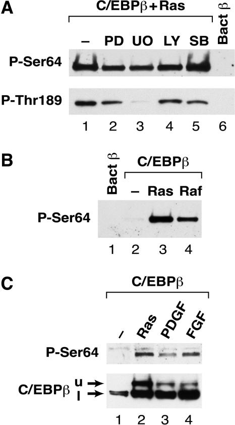
FIG. 4.
Analysis of the signaling pathway regulating Ser64 phosphorylation. (A) L cells were transfected with vectors for C/EBPβ and RasV12 (lanes 1 to 6) and then treated for 16 h in serum-free medium containing no inhibitor (lane 1) or the following inhibitors: 20 μM PD98059 (lane 2), 5 μM U0126 (lane 3), 50 μM LY294002 (lane 4), and 10 μM SB203580 (lane 5). Whole-cell lysates were prepared and analyzed by Western blotting using anti-P-Ser64 (upper panel) or anti- P-Thr189 (lower panel) antibodies. Bacterial C/EBPβ (lane 6) was included as a negative control. (B) Activated Raf-1 induces Ser64 phosphorylation. Cells were transfected with C/EBPβ alone (lane 2) or with RasV12 (lane 3) or constitutively active Raf-1 (lane 4). Cell extracts were analyzed by Western blotting using the anti-P-Ser64 antibody. Bacterial C/EBPβ was included as a control (lane 1). (C) PDGF and bFGF induce phosphorylation on Ser64. L cells were transfected with C/EBPβ, starved for serum overnight, and then either left untreated (lane 1) or stimulated for 4.5 h with 50 ng of PDGF/ml (lane 3) or 50 ng of bFGF/ml (lane 4). C/EBPβ coexpressed with RasV12 was included as a control (lane 2). Extracts were prepared and analyzed by Western blotting using anti-P-Ser64 (upper panel) and an antibody recognizing total C/EBPβ (lower panel). The upper (u) and lower (l) C/EBPβ species are indicated.

