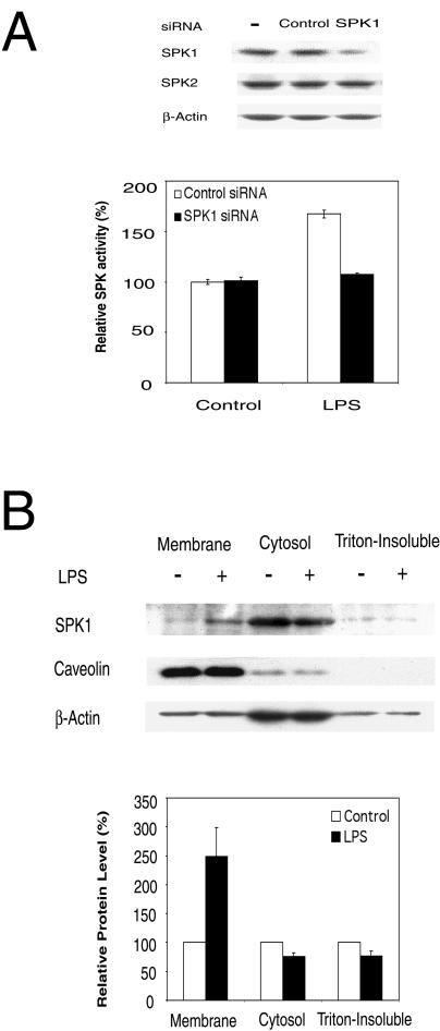
FIG. 2.
SPK1 is the major form of SPKs activated by LPS. (A) SPK1-specific siRNA blocks LPS-induced SPK activation. The upper panel shows that mouse SPK1 siRNA specifically down-regulated the expression of SPK1 but not SPK2 in RAW 264.7 cells. RAW 264.7 cells were treated with 175 nM siRNA for 48 h, and then cell lysates were subjected to Western blot analysis. The lower panel shows that SPK1 siRNA inhibited the activation of SPK by LPS. RAW 264.7 cells were transfected with 175 nM SPK1 siRNA for 24 h, serum starved overnight, and then treated with 10 ng of LPS/ml for 40 min. The SPK assay was conducted. (B) LPS-induced membrane translocation of SPK1. RAW 264.7 cells were treated in the absence or presence of 1 μg of LPS/ml for 1 h. Cell lysates were then fractioned into cytosol, membrane, and triton-insoluble membrane extracts by ultracentrifugation. Equal amounts of protein (30 μg) from each fraction were subjected to Western blot analysis (top panel). The band densities on the Western blot were analyzed by a densitometer, and relative ratios between LPS-treated and control results were reported. The value for controls was arbitrarily set to 100% (the bottom panel).

