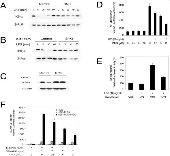
FIG. 5.
SPK mediates LPS-induced IκB-α degradation and NF-κB activation. (A) LPS-induced IκB-α degradation was inhibited by DMS in RAW 264.7 cells. RAW 264.7 cells were pretreated with or without 10 μM DMS for 30 min and then stimulated with 5 ng of LPS/ml for the indicated time course. IκB-α levels in cell lysates were detected by Western blotting. (B) LPS-induced IκB-α degradation was inhibited by SPK1 siRNA. RAW 264.7 cells were treated with 175 nM siRNA for 40 h and then stimulated with LPS (2 ng/ml) for the indicated time course. The IκB-α level was detected as described for panel A. (C) LPS-induced IκB-α degradation was inhibited by DMS in primary HMs. HMs were pretreated with or without 10 μM DMS for 30 min and then stimulated with 10 ng of LPS/ml for 20 min. IκB-α level was detected as described for panel A. (D) DMS inhibited NF-κB activation by LPS. RAW 264.7 cells were transiently transfected with Igκ-Luc along with a pEF-β-Gal vector. About 2 days after transfection, cells were stimulated with LPS (10 ng/ml) in the presence of indicated concentrations of DMS for 10 h. Reporter luciferase activity was measured using the Luciferase Assay system (Promega). Data were normalized with β-Gal activity in each sample. (E) DN-SPK1 inhibited LPS-induced NF-κB activation. RAW 264.7 cells were transfected with Igκ-Luc, pEF-β-Gal, and either a control vector or DN-SPK1. The reporter assay was conducted as described for panel D. (F) DMS only inhibited LPS-induced NF-κB activation in HEK 293 cells overexpressing both TLR4 and MD2. HEK 293 parental cells (HEK), HEK 293 cells overexpressing TLR4 (HEK-TLR4), and HEK 293 cells overexpressing both TLR4 and MD2 (HEK-TLR4/MD2) were plated on a 48-well plate, grown to confluence, and serum starved overnight. All three cell lines were also stably transfected with an NF-κB-dependent ELAM-1 luciferase reporter plasmid (pELAM-luc). Cells were stimulated with 100 ng of LPS/ml and 250 ng of recombinant human CD14/ml for 18 h. Reporter luciferase activity was measured using the Luciferase Assay system (Promega).

