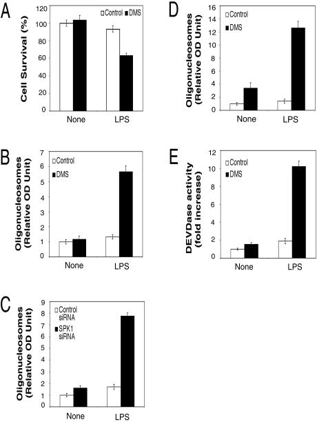
FIG. 6.
DMS and SPK1 siRNA sensitized LPS-activated macrophages to apoptosis. (A) DMS decreased cell survival in LPS-activated RAW 264.7 cells. RAW 264.7 cells were pretreated with 5 μM DMS for 30 min and then treated with 10 ng of LPS/ml for 24 h. Cell viability was measured by the MTT assay. (B) DMS sensitized LPS-activated RAW 264.7 cells to apoptosis. RAW 264.7 cells were pretreated with 5 μM DMS for 30 min and then treated with 10 ng of LPS/ml for 24 h. Apoptotic cell death was measured with a quantitative sandwich-ELISA assay detecting mono- and oligonucleosomes. The relative values for optical density at 405 nm minus that at 490 nm are reported. The value for untreated control was arbitrarily set to 1. (C) SPK1 siRNA sensitized LPS-activated RAW 264.7 cells to apoptosis. RAW 264.7 cells were transfected with control or SPK1 siRNA. One day after transfection, cells were pretreated with 5 μM DMS for 30 min and then treated with 10 ng of LPS/ml for 24 h. The apoptotic cell death was assayed as described for panel B. (D) DMS sensitized LPS-activated primary HMs to apoptosis. HMs were pretreated with 6 μM DMS for 30 min and then treated with 10 ng of LPS/ml for 24 h. The apoptotic cell death was assayed as described for panel B. (E) DMS enhanced LPS-induced caspase-3 activation. RAW 264.7 cells were pretreated with 5 μM DMS for 30 min and then treated with 10 ng of LPS/ml for 20 h. Then, the caspase-3 activities in cell lysates were assayed by measuring the hydrolysis of acetyl-Asp-Glu-Val-Asp p-nitroanilide. The relative values for optical density at 405 nm are reported.

