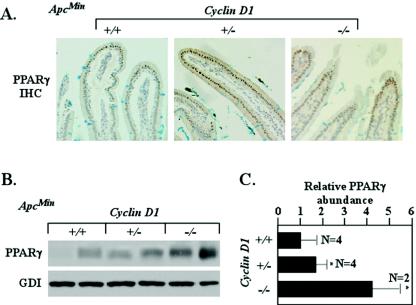
FIG. 5.
Genetic cyclin D1 deficiency increases PPARγ abundance in ApcMin intestinal epithelium. (A) Immunohistochemical (IHC) staining of duodenal epithelia of ApcMin mice. (B) Representative Western blot analysis of PPARγ1 in colonic epithelium in ApcMin mice (comparisons were made with 10 animals [4 cyclin D1+/+, 4 cyclin D1+/−, and 2 cyclin D1−/−] for each cyclin D1 genotype, two representative examples are shown for each genotype, and each lane represents lysate preparations made from individual animals with GDI as a loading control). (C) Analysis of mean PPARγ1 protein levels by densitometry (wt set to 1; mean and standard error of the mean; 13 weeks), normalized to GDI loading control. Asterisks indicate P values <0.05.

