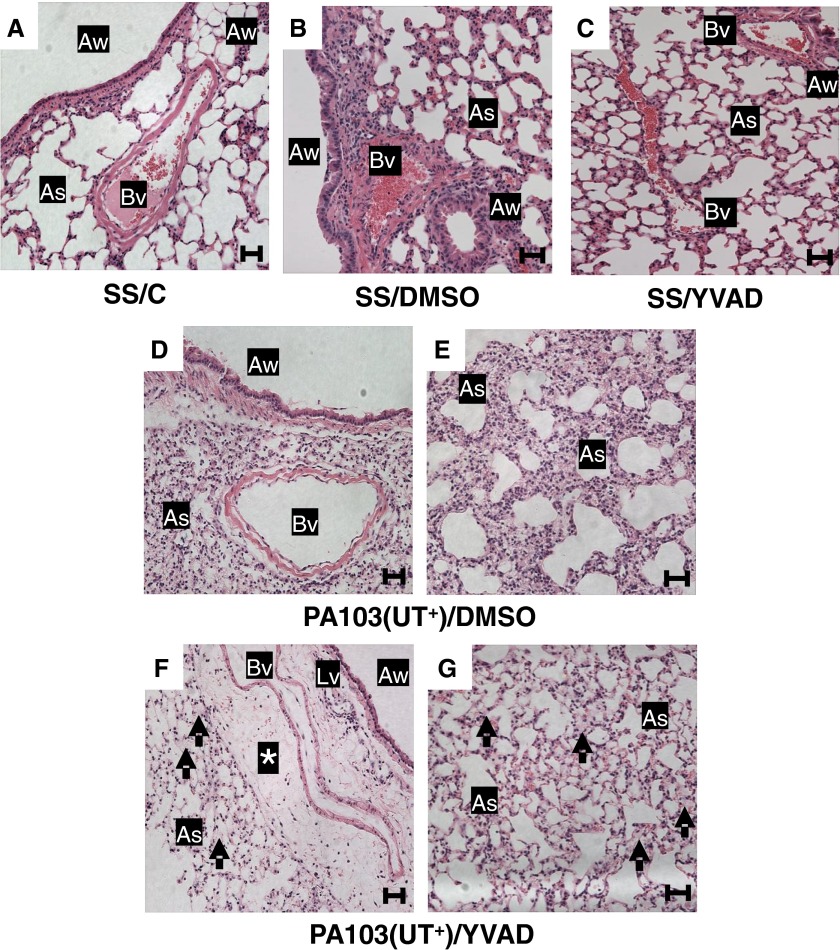
Figure 2.
Caspase-1 inhibition in P. aeruginosa–infected mice increases pulmonary edema. Groups of mice were inoculated intratracheally and injected intraperitoneally, as described in the legend to Figure 1. At 24 hours after inoculation, lungs were harvested under identical conditions, whereby the trachea was tied-off at the peak of inspiration, and the lungs removed and immersed in 10% neutral buffered formalin solution for 48 hours. Subsequently, paraffin-embedded sections were prepared for hematoxylin and eosin staining, and tissues analyzed by light microscopy (20× objective). For all groups, n = 3 mice. Representative images from each group are shown. (A) SS/C control group lungs showed normal architecture, no inflammation, and open airspaces. (B) SS/DMSO control group lungs showed normal architecture, no inflammation, and open airspaces. (C) SS/YVAD control group lungs showed normal architecture, no inflammation, and open airspaces. (D and E) PA103(UT+)/DMSO experimental group lungs from two different fields of view showing the bronchioarterial bundle (D) and the septal network (E). Sections displayed septal thickening and substantial cell infiltration. (F and G) PA103(UT+)/YVAD experimental group lungs from two different fields of view showing the bronchioarterial bundle (F) and the septal network (G). Sections displayed fluid accumulation in perivascular cuffs (asterisk) and fluid accumulation in air spaces (arrows) with a marked decrease in the number of infiltrating cells when compared with PA103(UT+)/DMSO experimental group lungs (D and E). Scale bars: 50 μm. As, air space; Aw, airway; Bv, blood vessel; Lv, lymphatic vessel.

