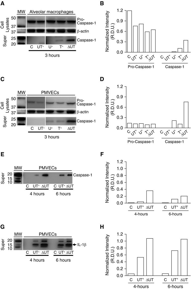
Figure 5.
Caspase-1 is activated in cultured rat pulmonary microvascular endothelial cells (PMVECs) in response to P. aeruginosa infection, and exoenzyme effectors ExoU and ExoT delay onset of caspase-1 activation. Rat alveolar macrophages or PMVECs growing in culture were untreated or independently inoculated with different P. aeruginosa strains at a multiplicity of infection (MOI) of 40 bacteria per host cell. At indicated time points after inoculation, cell lysates and clarified culture supernatants (“Super”) were prepared and analyzed by Western blot. Cell lysates were probed with an antibody against caspase-1 and subsequently stripped and reprobed with an antibody against β-actin. Clarified culture supernatants were probed with an antibody against caspase-1 or an antibody against IL-1β. (A) Effects of P. aeruginosa infection on caspase-1 activation in macrophages at 3 hours after inoculation. Macrophages were untreated (C) or inoculated with wild-type PA103(UT+), mutant PA103(U+) expressing only ExoU, mutant PA103(T+) expressing only ExoT, or double-mutant PA103(ΔUT) expressing no exoenzyme effectors. The top panels show expression of the inactive pro–caspase-1 enzyme in whole-cell lysates. The middle panels show β-actin levels as a loading control. The bottom panels show activated caspase-1 released to the culture medium. MW, molecular weight markers. (B) Representative densitometric analysis of macrophage pro–caspase-1 was normalized to β-actin for cell lysates. Active caspase-1 was normalized to sample volume. Data are expressed as relative density units (R.D.U.). (C) Effects of P. aeruginosa infection on caspase-1 activation in PMVECs at 3 hours after inoculation under the untreated and infection conditions described for A above. (D) Representative densitometric analysis of PMVEC pro–caspase-1 was normalized to β-actin for cell lysates. Active caspase-1 was normalized to sample volume. (E) Effects of P. aeruginosa infection on caspase-1 activation 4 and 6 hours after inoculation in PMVECs that were untreated (C) or inoculated with either wild-type PA103(UT+) or double-mutant PA103(ΔUT). (F) Representative densitometric analysis of PMVEC active caspase-1 was normalized to sample volume. (G) Effects of P. aeruginosa infection on IL-1β activation 4 and 6 hours after inoculation in PMVECs that were untreated (C) or inoculated with either wild-type PA103(UT+) or double-mutant PA103(ΔUT). (H) Representative densitometric analysis of PMVEC active IL-1β was normalized to sample volume. (A–H); n = 3 experiments; representative blots and corresponding densitometry are shown.

