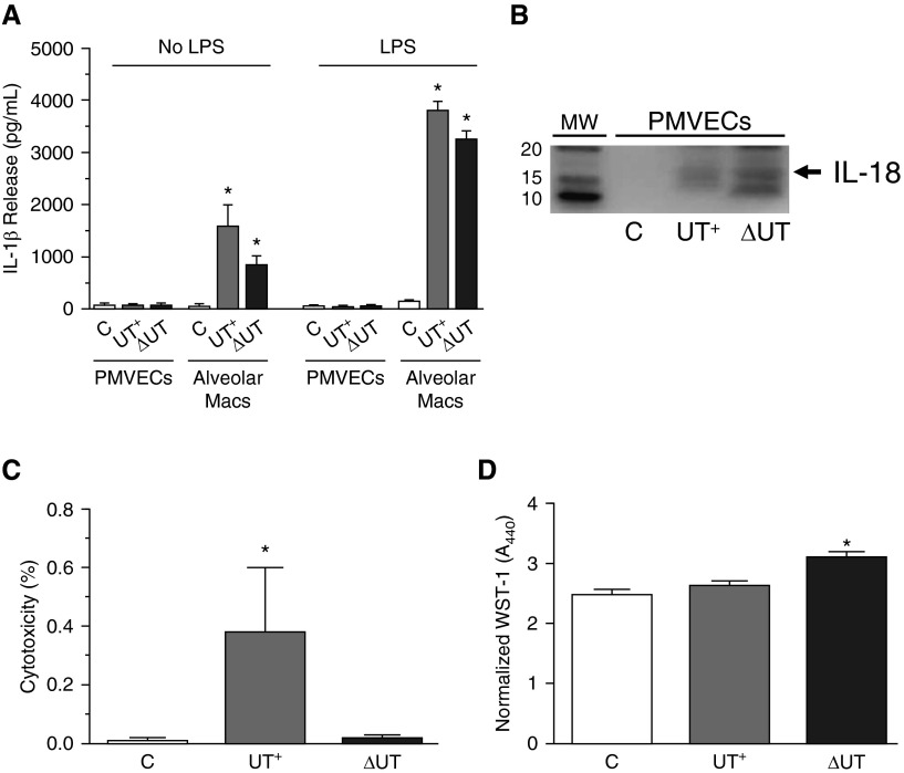
Figure 6.
Caspase-1 activation in PMVECs results in modest IL-1β and IL-18 activation and does not induce pyroptotic cell death. Rat alveolar macrophages or PMVECs growing in culture were untreated or independently inoculated with either P. aeruginosa PA103(UT+) or the isogenic double-mutant PA103(ΔUT) strain at a MOI of 40 bacteria per host cell. Cells were analyzed at 3 hours after inoculation. (A) Culture supernatants from control (C) PMVECs and macrophages were collected and IL-1β activation/secretion was measured by ELISA. Note that the absolute levels of IL-1β secretion by PMVECs were negligible, even in the presence of LPS pretreatment (100 ng/ml, overnight incubation), a critical priming signal known to increase IL-1β and inflammasome component gene expression in macrophages. (B) Clarified culture supernatants from control (C) or infected PMVECs were prepared and analyzed for IL-18 levels by Western blot. (C) Clarified culture supernatants from control (C) or infected PMVECs were analyzed for lactate dehydrogenase (LDH) release into the culture medium as an indicator of cell death. Data represent percent released as normalized to total LDH assayed in the cell monolayer. (D) Control (C) or infected PMVECs were analyzed for metabolism of WST-1 as an indicator of cell viability. Normalized WST-1 conversion was measured spectrophotometrically at A440nm after 1 hour of incubation and corrected for background at A620nm. (A–D) n = 3 experiments. All data represent mean ± SEM. *P < 0.05 by one-way ANOVA (Newman-Keuls post hoc test) when compared within the group. Macs, macrophages; WST-1, 4-[3-(4-iodophenyl)-2-(4-nitro-phenyl)-2H-5-tetrazolio]-1,3-benzene disulfonate.

