Figure 5.
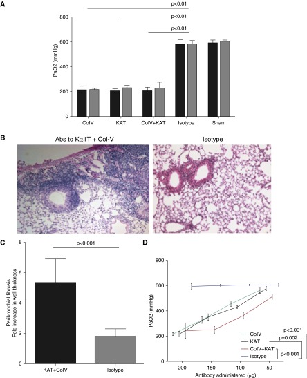
Preexisting lung-restricted antibodies induce allograft dysfunction and prevent development of tolerance. Wild-type donor BALB/c lungs were transplanted into C57BL/6 recipients. A combination of MHC-related 1 and cytotoxic T-lymphocyte–associated protein 4-Ig was used to induce tolerance. We administered Col-V (200 μg), KAT (200 μg), Col-V plus KAT (200 μg each), or isotype control antibodies on Days −2, −1, 0, 6, 7, and 8, and weekly thereafter. (A) On Days 30 (black bars) and 45 (gray bars) arterial blood gases were obtained to analyze allograft function. Sham mice underwent ventilation for 1 hour without lung transplantation. (B and C) Histology revealed developement of peribronchial fibrosis in mice treated with lung-restricted antibodies compared with isotype control. For calculation of peribronchial fibrosis, the average of maximum bronchial wall thickness in 10 high-power fields in mice within each treatment group was divided by average wall thickness in native lungs to determine fold increase. (D) The effects of lung-restricted antibodies were dose dependent. Mice underwent serial administration of lung-restricted antibodies at the time points described previously here. On Day 45, allograft function was analyzed by arterial blood gases. Data are presented as mean (±SEM).

