
Fig. 1.
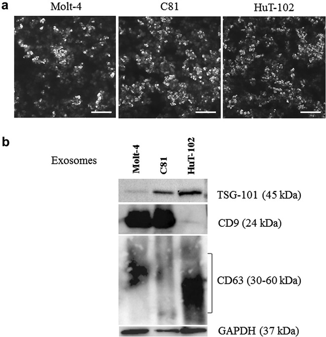
Characterization of exosomes derived from leukemic cells. a Scanning electron micrograph of exosomes isolated from Molt-4, C81 and HuT-102 cells, scale bar 1 µm. b A representative western blot showing the protein expression of TSG-101, CD9, CD63 and GAPDH in the lysates of exosomes (15 µg) isolated from leukemic cells