
Fig. 4.
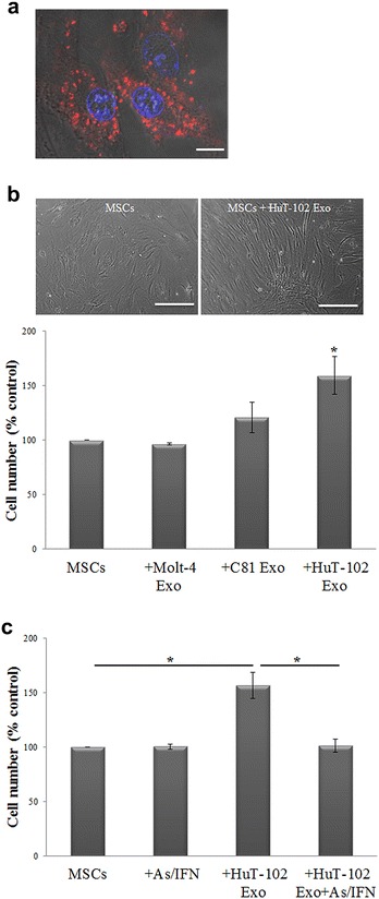
Induction of MSCs proliferation by ATL-derived exosomes. a Fluorescence images of MSCs following the uptake of PKH26-labeled HuT-102 exosomes (red color), scale bar 10 µm. Hoechst is used as a nuclear stain (blue color). b Bright field images of MSCs cultured alone or with 30 µg of HuT-102-derived exosomes for 72 h, scale bar 40 µm. Histogram representing the proliferation of MSCs after co-culture with leukemia-derived exosomes for 72 h (*p < 0.05). c Histogram representing the proliferation of MSCs after co-culture with HuT-102-derived exosomes for 72 h, with or without As/IFN treatment (*p < 0.05)