
Figure 8.
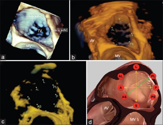
Tricuspid annulus positioned in the surgical view as: three-dimensional volumetric data (a), an Image Arena reconstruction (b), Image Arena reconstruction with increased transparency (c), and a simulation using HeartWorks (Inventive Medical Limited, London, UK) software (d). The HeartWorks image is annotated with the observed axis of maximal dynamism (green) and the axes routinely measured during imaging (yellow) (A: Anterior, AL: Anterolateral, AS: Anteroseptal, AV: Aortic valve, L: Lateral, MV: Mitral valve, P: Posterior, PL: Posterolateral, PS: Posteroseptal, S: Septal, TV: Tricuspid valve)