
Fig. 2.
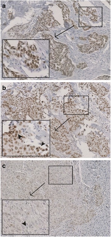
Immunohistochemical PARP1 staining in TNBC tissue microarrays. a b BRCA1-like TNBC with high (3+) nuclear PARP1 levels in tumor cells (10× magnification) as assessed by a pathologist. 3+ stained nuclei are exemplarily indicated by black arrows in a separate image section. c Non-BRCA1-like TNBC with low cytosolic and nuclear PARP1 levels in tumor cells (10× magnification). Black arrow shows an unstained nucleus. Tissue microarrays were incubated with mouse anti-PARP antiserum followed by staining with peroxidase-conjugated secondary antibody molecules and diaminobenzidine (DAB+) as chromogenic substrate. Nuclear counterstaining was performed with hematoxylin