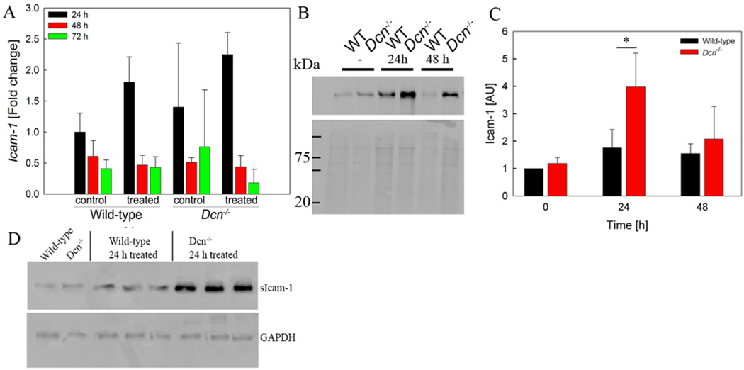
FIGURE 3. ICAM-1 expression is differentially altered in wild-type and Dcn−/− mice during DTH activity.
(A) Real-time PCR for ICAM-1 expression during the time course of DTH (P = n.s., n=3–5) (B,C) Increased Icam1 expression in Dcn−/− mouse tissues is confirmed by Western blotting 24h after DTH induction. (B) Representative Western blot. Ponceau S staining is used as loading control (lower panel) (C) Densitometric analysis (P = 0.039; n=4). (D) Western blot analysis of ICAM-1 expression in plasma of wild-type and Dcn−/− mice prior to and 24h after DTH elicitation. 20µg of protein/lane were analysed. GAPDH is used as a loading control.

