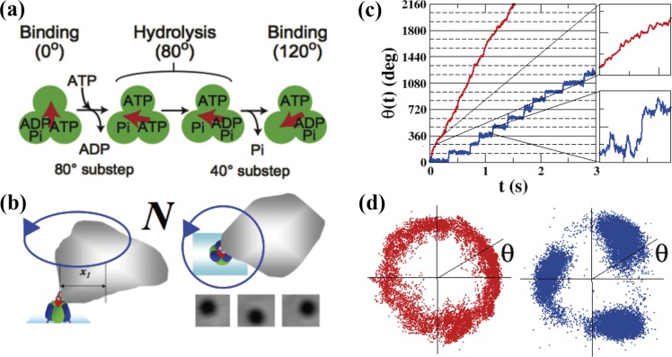
Figure 3.
Rotary motor protein, F1. (a) Reaction scheme of F1. The green circles and the red arrow represent the ß subunits and γ subunit of F1, respectively. F1 performs a 120° step rotation upon ATP hydrolysis consisting of 80° and 40° substeps. (b) Schematic diagrams of our experimental system (not to scale). The rotation of F1 is probed by an irregularly shaped magnetic bead (see Methods in Hayashi et al.11 for this irregular shape). The size of F1 is about 10 nm and the size of the bead is about 300–500 nm. (c) ATP-driven rotations of F1 probed by the magnetic beads at 1 mM ATP (red) and 100 nM ATP (blue). The recording rate was 2000 fps. (d) The center of mass of the bead was calculated from the recorded images for the case of 1 mM ATP (red) and 100 nM ATP (blue).

