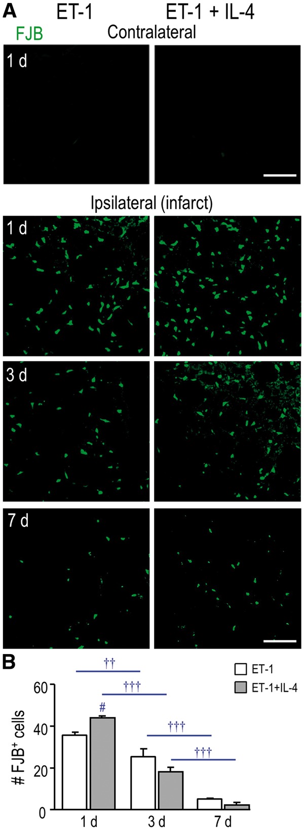
FIGURE 5.

IL-4 treatment did not rescue neurodegeneration in the infarct. (A) Representative images show FJB (green), which was used to identify degenerating neurons. The contralateral and ipsilateral striatum (infarct region) from the same rat are shown (either untreated or IL-4 treated) for 1 day. The ipsilateral infarct is also shown for 3 and 7 days. Scale bar = 100 μm and applies to all images. (B) Summary of the number of FJB-labeled cells per 200 µm2 in the infarct at 1, 3, and 7 days. The symbols represent: † time-dependent changes within a treatment group; # IL-4 effects. One symbol indicates p < 0.05; 2 symbols, p < 0.01; 3 symbols, p < 0.001.
