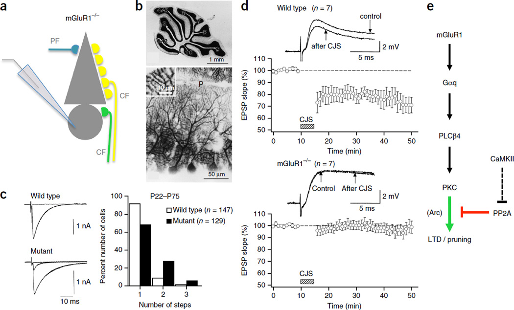
Figure 2.
Impaired CF synapse elimination and deficient PF LTD in mGluR1 knockout mice. (a) Schematic of the patch-clamp configuration used to record from an mGluR1 knockout Purkinje cell (PC) that is illustrated with multiple CF innervation. (b) Gross anatomy of the cerebellum (top, Nissl staining) and morphology of PC dendrites (bottom, calbindin immunostaining) are normal in mGluR1 knockout mice. (c) Persistent multiple CF innervation in adult mGluR1 knockout mice (P22–P75). CF-mediated EPSCs (left) and frequency distribution histogram showing the number of discrete CF EPSC steps at increasing stimulus strength (right), representing the number of CF inputs. (d) LTD at PF PC synapses is deficient in adult mGluR1 knockout mice. In wild-type mice, EPSPs elicited by PF stimulation undergoes LTD after conjunctive PF and CF stimulation (CJS) at 1 Hz for 5 min (top). In contrast, PF EPSPs are not depressed by CJS in mGluR1 knockout mice (bottom). All values are shown as mean ± s.e.m. (e) mGluR1 signaling pathway. CaMKII activation contributes to LTD through an indirect blockade of PP2A. CaMKII may similarly contribute to CF synaptic pruning, but this has not yet been verified. mGluR1, type 1 metabotropic glutamate receptor; Gαq, G-protein αq; PLCβ4, phospholipase Cβ4; PKC, protein kinase C; αCaMKII, α isoform of calcium/calmodulin-dependent kinase II; PP2A, protein phosphatase 2A; Arc, activity-regulated cytoskeleton-associated protein (also known as Arg3.1). Panels b,c adapted from ref. 73, Elsevier; d from ref. 47, AAAS.

