Figure 6.
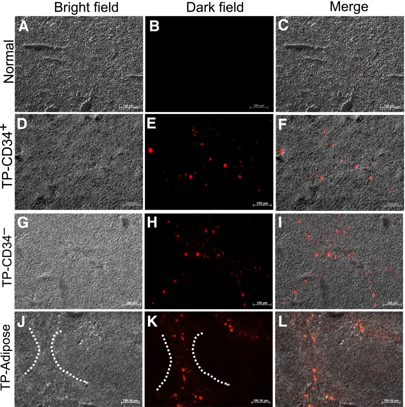
Engraftment of human mesenchymal stromal cells in the thioacetamide (TAA)-injured mice liver. The frozen sections of normal liver (A–C), CD34+ amnion membrane-derived stem/progenitor cells (AMSPC) liver (D–F), CD34− amnion membrane-derived stromal fibroblast cell (AMSFC) liver (G–I), and adipose tissue-derived mesenchymal stem cells (ADMSC) liver (J–L) were shown under a bright field (A, D, G, J), a fluorescent field (B, E, H, K), and a merged field (C, F, I, L). PKH26-labeled CD34+ AMSPCs, CD34− AMSFCs, and ADMSCs engrafted into TAA-injured mice tissues were shown in (E), (H), and (K). The dashed lines in (J) and (K) showed the exemplified bridging fibrosis area of the stem/progenitor cells homing. Scale bar = 100 μm. Abbreviation: TP, transplantation.

