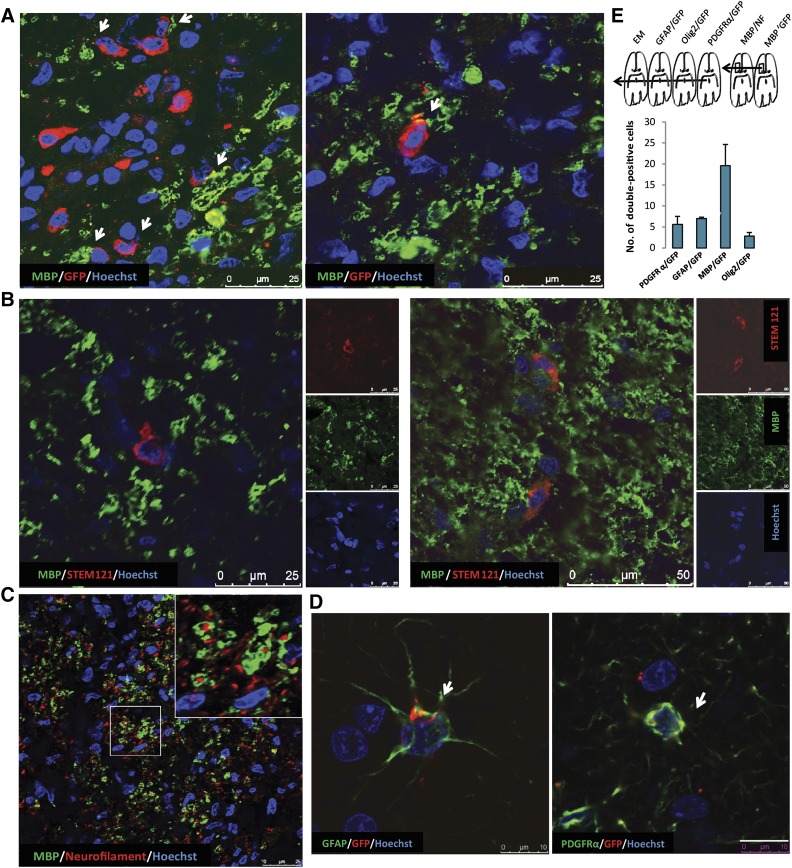
Figure 4.
Analysis of hiPSC-derived OPCs in EAE marmosets. (A): GFP/MBP double immunostaining indicates that the implanted cells are capable of producing myelin. Scale bars: 25 µm. (B): STEM121/MBP double immunostaining indicates that the implanted cells are capable of producing myelin. Scale bars: 25 µm. (C): Neurofilament/MBP double immunostaining in the same area shows the close interaction of MBP-expressing implanted cells with unmyelinated axons. (D): Likewise, GFP/PDGFRα immunostaining reveals immature OPCs (arrows) among the implanted cells as well as a few GFP/GFAP double-stained cells (arrows), suggesting differentiation of the implanted cells toward astrocytes. Scale bars: 50 µm. (E): Scheme indicating the order of subsequent marmoset brain sections used for immunohistochemical analyses and electron microscopy studies. Quantification of PDGFRα-, GFAP-, MBP-, and Olig2-positive implanted (GFP+) cells in one standard area within the corpus callosum at 40 days after implantation indicates that the majority of implanted cells became MBP-positive oligodendrocytes. Data presented as mean ± SEM. Abbreviations: EAE, experimental autoimmune encephalomyelitis; EM, standard error of the mean; GFAP, glial fibrillary acidic protein; GFP, green fluorescent protein; hiPSC, human induced pluripotent stem cell; MBP, myelin basic protein; NF, neurofilament; OPC, oligodendrocyte precursor cell, PDGFRα, platelet-derived growth factor receptor-α.

