
Figure 3.
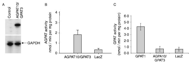
AGPAT enzymatic activity of wild-type human AGPAT10/GPAT3 in HEK-293 cells. (A) Shown is the western blot for the cell pool, used for determining the acyltransferase activity, for the V5 epitope-tagged AGPAT10/GPAT3 as probed with V5-antibody. GAPDH is included to demonstrate equal loading. (B) The enzymatic activity determined as conversion of 3H-LPA to 3H-PA in the presence of oleoyl-CoA, and expressed as product (3H-PA) formed per minute per mg protein. The LPA to PA conversion by recombinant adenovirus β-galactosidase (LacZ) was used as a control. Not shown is the conversion of substrate in the absence of enzyme. (C) Glycerol-3-phosphate acyltransferase enzymatic activity of AGPAT10/GPAT3 in the HEK-293 cell lysate. The GPAT activity was determined by incubating [14C]-glycerol-3-phosphate with an acyl-CoA. Recombinant adenovirus expressing human GPAT1 was included as a positive control. The recombinant adenovirus β-galactosidase (LacZ) was used as a control. Not shown is the conversion of substrate in the absence of enzyme.