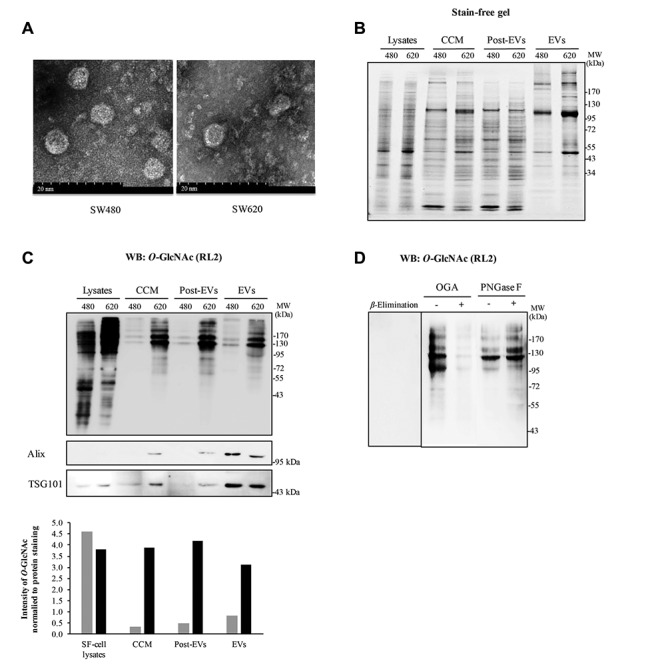
Figure 2. EVs isolated from conditioned medium of SW480 and SW620 cells containing O-GlcNAcylated proteins. A: Representative captured pictures using transmission electron microscopy revealing enriched EVs of SW480 and SW620 cells varied in size from 40-80 nm. Scale bar = 20 nm. B: Stain-free SDS-PAGE gel demonstrated the pattern of total proteins from cell lysates cultured in serum free-medium (SF-cell lysates) and different fractions of EV preparation steps (CCM, post-EVs and EVs). C: Detection of O-GlcNAcylated proteins from SF-cell lysates and different fractions of EV preparation steps by Western blotting of O-GlcNAc using RL2 antibody. Exosomal markers (Alix and TSG101) showed an enrichment of exosomes in the final fraction (EVs). D: Western blots of O-GlcNAc of EV proteins from SW620 cells treated with OGA, PNGase F and on-blot β- Elimination. Post-EVs refers to sucrose cushion fraction. 480 refers to SW480, while 620 refers to SW620.