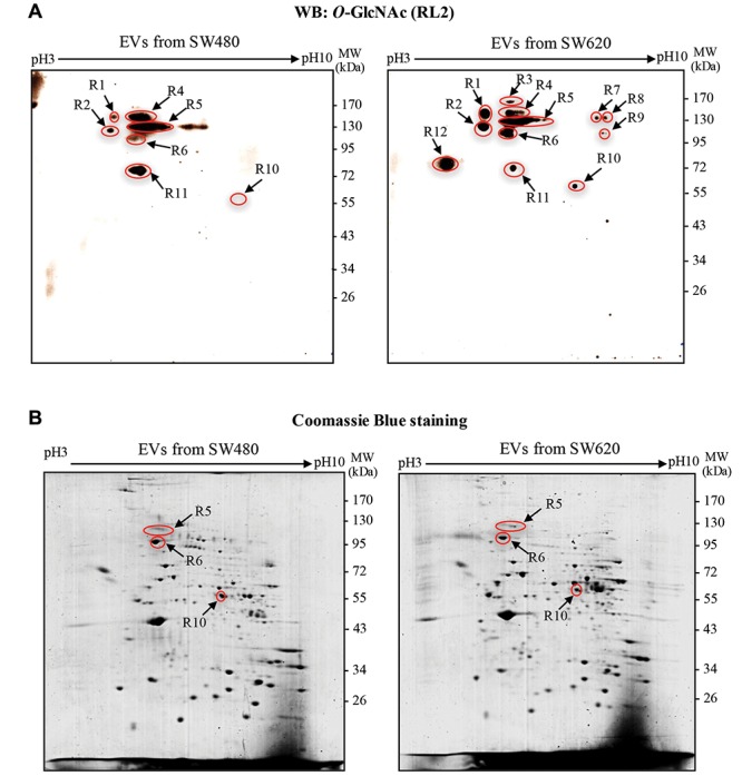
Figure 3. O-GlcNAc modification of EV proteins from SW480 and SW620 cells. A: Representative 2DE immunoblots of O-GlcNAcylated EV proteins isolated from SW480 and SW620 cells. Arrows indicate O-GlcNAc-protein spots. B: Representative 2DE gels of EV proteins from SW480 and SW620 cells. Total protein spots were visualized by staining the gels with Coomassie Brilliant Blue R250. Arrows indicate protein spots that could be matched with O-GlcNAc immunoblots. WB, Western blot.