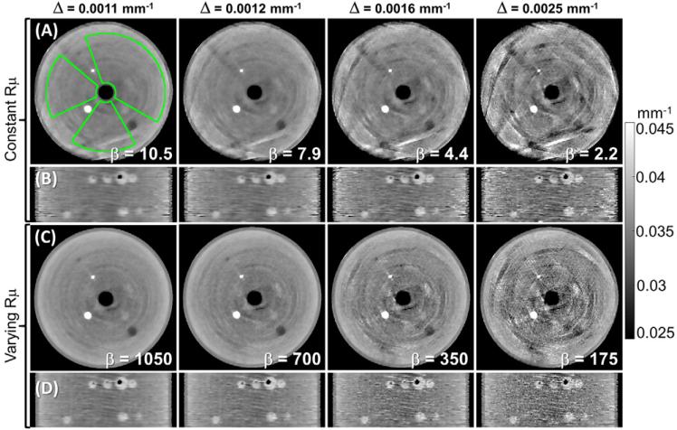
Figure 6.
Images of the gelatin phantom reconstructed with various levels of regularization strength. (A), (B) Spatially constant and (C), (D) spatially varying regularization. The non-uniformity Δ was measured in the ROI shown in (A). Columns correspond to images at matched Δ. The spatially varying penalty achieved reduction in artifacts, sharper edges, and superior noise and resolution homogeneity than the spatially constant penalty at matched Δ.

