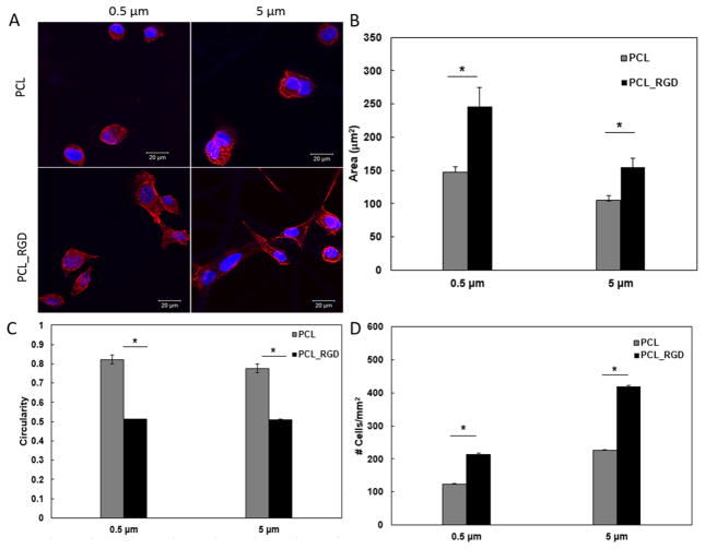
Fig. 4.
Characterization of the initial attachment and spreading of MDCK cells on small and large diameter fibers, with or without RGD conjugation. (A): Confocal images of MDCK cells cultured on PCL-based scaffolds (40x). Red: F-actin; Blue: nuclei. (B): Cell spreading as a function of fiber diameter and surface chemistry; (C): Cell circularity as a function of fiber diameter and surface chemistry. The circularity is defined as 4π×area/perimeter2. (D): Average number of cells attached to the scaffold per mm2 surface area. Quantification was carried out using ImageJ software based on five separate 1,024 × 1,024 μm2 confocal images. *Significantly different (p<0.05, ANOVA) from RGD conjugated scaffolds. No significant difference was observed between small and large diameter fibers (p>0.05). Error represents standard error of the mean of 3 repeats.

