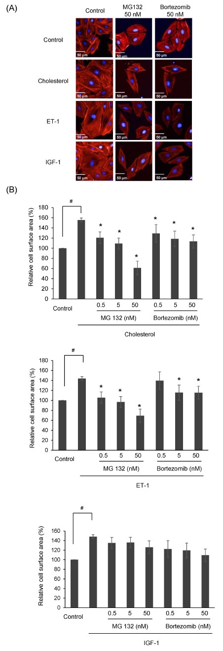
Fig. 1. Effects of proteasome inhibitors on cell surface area of cholesterol-induced cardiac hypertrophic H9c2 cells. (A) H9c2 cells were cultured in serum-free medium for 4 h and treated with MG132 (0.5-50 nM) or Bortezomib (0.5-50 nM) in the presence of ET-1 (100 nM), IGF-1 (50 ng/ml), and cholesterol (5 μg/ml) for 24 h. Cells were fixed, and then stained with rhodamine phalloidin (red) and DAPI (blue) for 30 min to visualize F-actin and nuclei, respectively. (B) Images were acquired on an Operetta system (PerkinElmer, USA) and the cell surface area was analyzed using HarmonyⓇ High Content Imaging and Analysis Software. The values shown are the mean ± S.D. from three independent experiments. #P < 0.05 vs control; *P < 0.05 vs cholesterol or ET-1.

