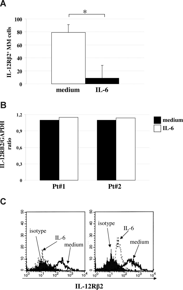
Figure 3.

Down-regulation of IL-12Rβ2 by IL-6. (A) Down-regulation of IL-12Rβ2 expression in primary MM cells on incubation with medium (□) or IL-6 (■) for 48 hours, as assessed by flow cytometry using the Santa Cruz antibody. Results represent median IL-12Rβ2+ cells plus or minus SE from 4 different experiments. (B) Quantitative analysis of IL-12RB2 versus GAPDH transcript in 2 MM cell suspensions (patients 1 and 2) cultured with (□) or without (■) IL-6. (C) IL-12Rβ2 surface expression in normal PPCs before (open profile) and after (dashed line) treatment for 48 hours with hrIL-6, as assessed by flow cytometry using the BD Bioscience mAb. Dark profile indicates staining with isotype-matched mAb. Two different experiments of the 4 performed with superimposable results are shown.
