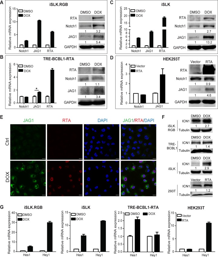
Fig 1. RTA up-regulates Notch components in different cell types.
(A–C) iSLK.RGB cells, TRE-BCBL1-RTA cells, and iSLK cells were plated in 6-well plates at 0.1 million cells per well. After 24 h, the cells were induced with doxycycline (5 μg/ml) for 24 h and the expression of Notch components was measured by qPCR and western blotting. The data were normalized to GAPDH expression. (D) HEK293T cells seeded in 6-well plates were transfected with RTA (4 μg) using Lipofectamine 2000 for 24 h and the expression of Notch components was measured by qPCR and western blotting. (E) Immunofluorescence imaging of iSLK cells treated with or without doxycycline for 24 h. The JAG1 (Green) and RTA (Red) in the nucleus were labeled with the indicated primary and secondary antibodies. Scale bars represent 20 μm. (F) iSLK.RGB cells, TRE-BCBL1-RTA cells and iSLK cells were plated in 6-well plates at 0.1 million cells per well and treated with or without doxycycline for 24 h. HEK293T cells were transfected with RTA (4 μg) using Lipofectamine 2000 for 24 h in 6-well plates. The protein level of ICN1 was quantified by western blotting. (G) Hes1 and Hey1 were quantified by qPCR. The data were normalized to GAPDH expression. Data were expressed as the mean ± s.e.m., n = 3, *p<0.05.

