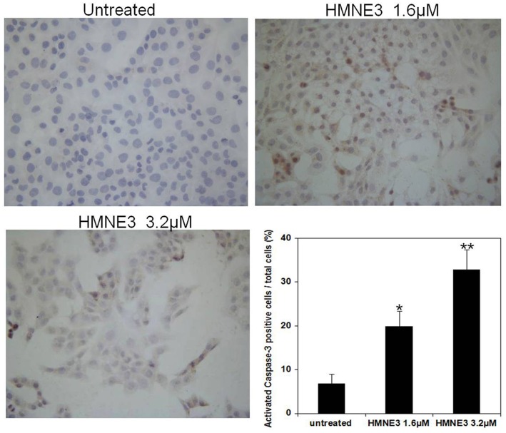
Fig 6. Immunocytochemical staining of activated Caspase-3 in Capan-1 cells following HMNE3 treatment.
Approximately 2×10⁴ cells were plated on coverslips that were pre-placed in 12-well plates. All of the cells were treated with the indicated doses of HMNE3 for 48 h. The cells were fixed and incubated with 5% BSA for 30 min. The cells were incubated with a rabbit anti-human cleaved Caspase-3 antibody overnight at 4°C, washed with PBS, and incubated with a biotinylated horse anti-rabbit IgG antibody for 30 min. After rinsing with PBS for 5 min, streptavidin-horseradish peroxidase enzyme complex was diluted in PBS and incubated with the cells for 30 min. After washing with PBS, the staining was visualized by incubating the cells in DAB and counter-stained with a hematoxylin solution. The cells were imaged using a microscope. * P<0.05 compared with the control group; Student’s t test.

