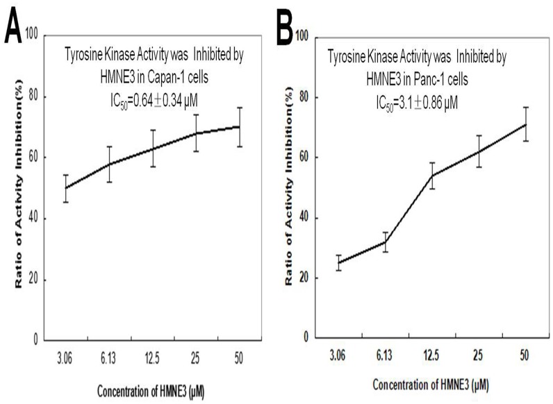
Fig 7. HMNE3 inhibited tyrosine kinase activity in the Capan-1 (A) and Panc-1 cells (B).
Ninety-six-well plates were pre-coated with 20 μg/mL of a 4:1 poly (Glu,Tyr) solution as a substrate. An ATP solution was added to each well. Varying doses of HMNE3 were added to each test well. The kinase reaction was initiated by the addition of purified tyrosine kinase proteins diluted in 49 μL of kinase reaction buffer. After incubation for 1 h at 37°C, the plate was washed thrice with PBST. An anti-phosphotyrosine (PY99) antibody was then added. After a 30 min incubation at 37°C, the plate was washed thrice, and 100 μL of a horseradish peroxidase-conjugated goat anti-mouse IgG was added. The reaction was terminated by the addition of 50 μL of 2 mol/L H2SO4, and the plate was read at 490 nm using a multi-well spectrophotometer.

