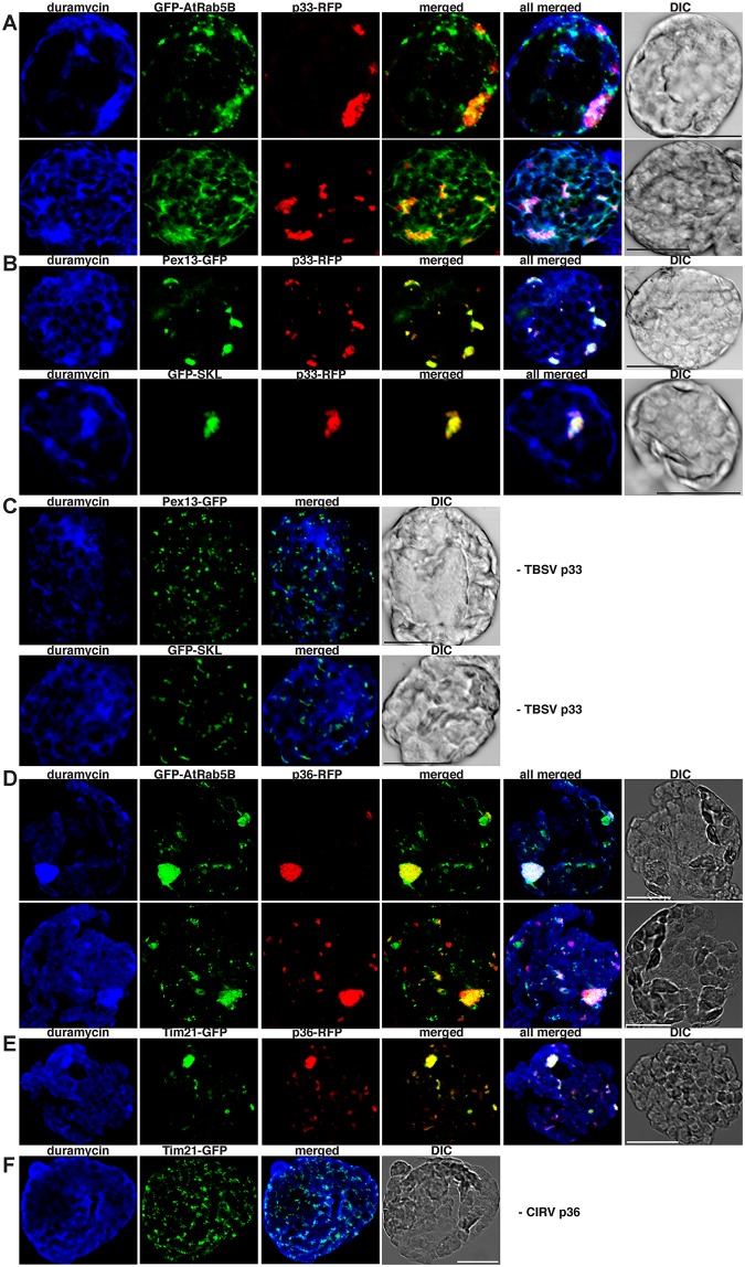
Fig 4. Rab5 is partly co-localized with PE-enriched tombusvirus replication compartment in plant cells.
(A) Confocal laser microscopy images show the co-localization of GFP-AtRab5B with the TBSV p33-RFP replication protein in subcellular areas enriched with PE in N. benthamiana protoplasts. Scale bars represent 20 μm in each panel. (B) Confocal laser microscopy images confirm that these subcellular areas are derived from aggregated peroxisomes based on co-localization with either Pex13-GFP peroxisomal membrane marker protein or GFP-SKL peroxisomal luminal marker protein. (C) Control images show the lack of PE enrichment in peroxisomes in the absence of viral components. Note the absence of aggregated peroxisomes in these cells. (D) Confocal laser microscopy images show the co-localization of GFP-AtRab5B with the CIRV p36-RFP replication protein in subcellular areas enriched with PE. (E) Confocal laser microscopy images confirm that these subcellular areas are derived from aggregated mitochondria based on co-localization with Tim21-GFP marker protein. (F) Control images show the lack of PE enrichment in mitochondria in the absence of viral components. Note the absence of aggregated mitochondria in these cells.

