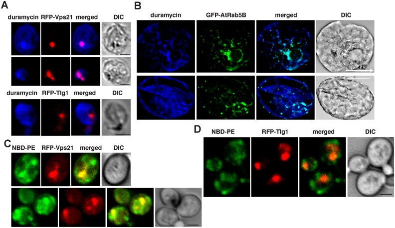
Fig 5. The early endosomal membranes are enriched with PE in yeast and plant cells.
(A) Confocal laser microscopy images show the enrichment of PE and its co-localization with the early endosomal RFP-tagged Vps21p expressed from TEF1 promoter in the absence of tombusviral components in wt yeast cells (top two images). DIC images are shown on the right. Localization of PE is detected by using biotinylated duramycin peptide and streptavidin conjugated with Alexa Fluor 405. The bottom image shows the lack of PE enrichment in trans-Golgi network marked by RFP-Tlg1. Scale bars represent 2 μm. (B) Confocal laser microscopy images show the enrichment of PE with the endosomal GFP-tagged AtRab5B expressed from 35S promoter in the absence of tombusviral components in N. benthamiana cells. Scale bars represent 20 μm. (C) Enrichment of exogenous PE in early endosomes labeled with RFP-Vps21 protein in wt yeast cells. Yeast cells were cultured (initial 0.3 OD600) with 80 μM NBD-PE for 12–14 h. Scale bars represent 2 μm. (D) The control panel shows minimal level of NBD-PE enrichment in the trans-Golgi network labeled with RFP-Tlg1 in wt yeast cells. Scale bars represent 2 μm.

