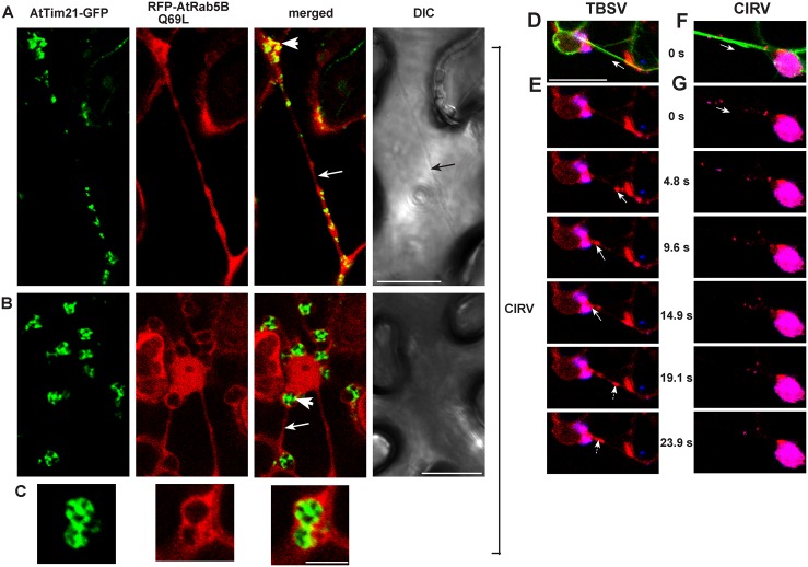
Fig 6. The role of actin filaments in recruitment of Rab5-positive endosomes to the large tombusviral replication compartments in plant cells.
(A–B) Confocal laser microscopy images of CIRV-infected N. benthamiana cells expressing AtTim21-GFP mitochondrial marker and the RFP-tagged active GTP-locked AtRab5B mutant. Note the large aggregated mitochondria-containing area (marked by a white arrowhead) and the actin-like filamentous structure (pointed at by a white arrow). Scale bars represent 20 μm. (C) An enlarged subcellular area showing the aggregated mitochondria and the RFP-AtRab5 mutant. Scale bar represents 5 μm. (D–G) Still images from a movie taken from plant cells co-expressing RFP-AtRab5B with TBSV p33-BFP (D–E) or CIRV p36-BFP (F–G) in transgenic plants expressing GFP-mTalin (an actin filament marker). Scale bars represent 20 μm. (D and F) All three channels from 0s are shown. White arrow depicts the direction of Rab5-positive endosomes (red) moving towards the replication compartment (blue) via actin filaments (green). (E and G) Merged images of RFP-AtRab5B and p33-BFP/p36-BFP. White arrow shows the movement of Rab5-positive endosomes. Scale bars represent 20 μm. See S2 and S3 Videos.

