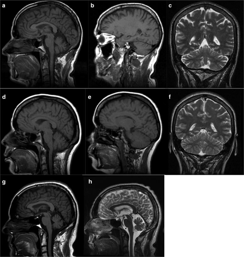
Figure 2.
Brain MRI of FC cases with SPG7 variants. (a–f) Sagittal T1-weighted (a, b, d, e) and coronal T2-weighted (c, f) images show moderate atrophy of the cerebellar vermis and hemispheres in subject 2 at age 57 (a–c), whereas cerebellar atrophy is milder in subject 3 at age 51 (d–f). Sagittal T1- (g) and T2- (h) weighted images show mild cerebellar atrophy on the initial MRI of subject 13 at age 51 (g) without significant progression at the follow-up MRI at age 58.

