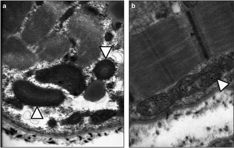
Figure 1.
Electron microscopy of patient muscle indicates abnormalities in mitochondria. (a) Electron micrograph of patient muscle with arrowheads indicating mitochondria with abnormal, concentric cristae. (b) Electron micrograph of patient muscle with arrowhead indicating mitochondria with increased dark, granular inclusions.

