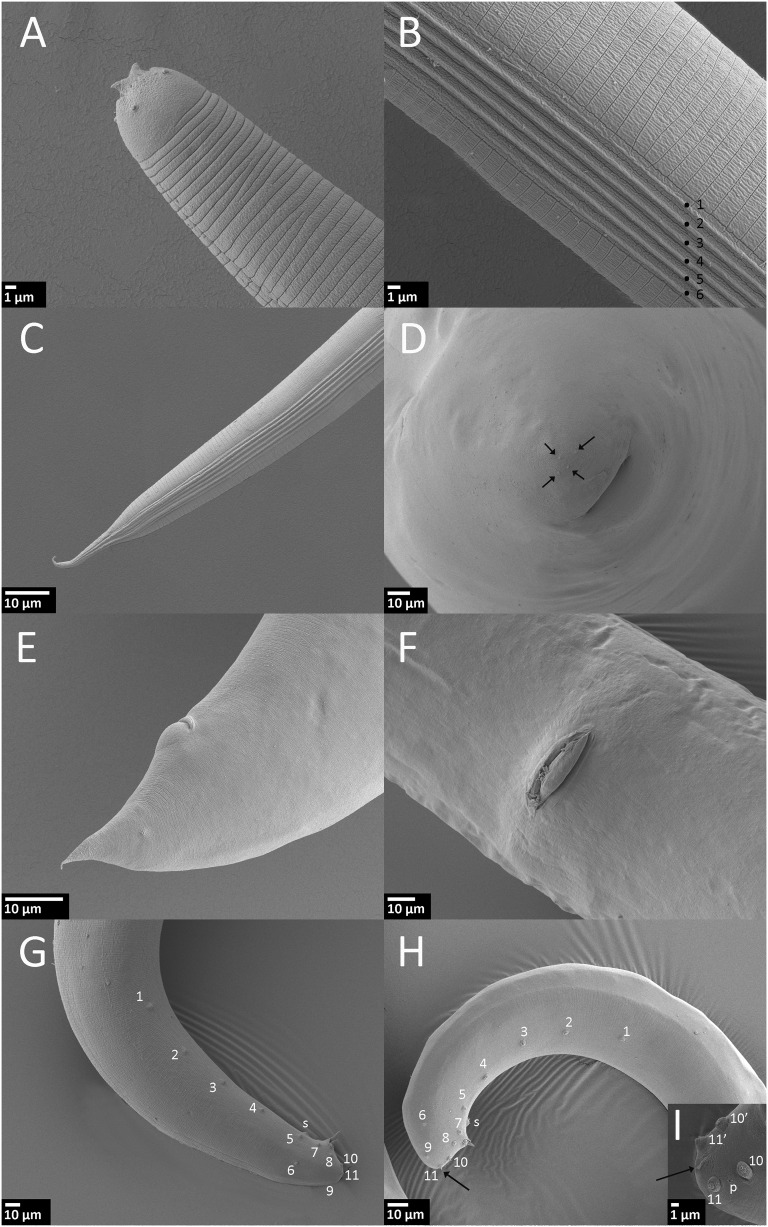
Fig. 2.
Steinernema biddulphi n. sp. Scanning electron microscopy of infective juvenile (IJ), male and female. A–C. IJ. A. Head region with horn-like structures. B. Lateral field in mid-body (ridges numbered 1 to 6). C. Lateral field in tail region. D. First generation female, tail, and four projections on tip of the tail (arrow). E, F. Second generation female. E. Tail with postanal swelling. F. Vulva. G. First generation male, tail with paired genital papillae (numbered) and single papilla (s), lateral. H, I. Second generation male. H. Tail with paired genital papillae, single papilla (s) and mucron (arrow). I. Tail with a part of paired genital papilae (numbered), mucron (arrow), and phasmid opening (p), ventro-lateral.

