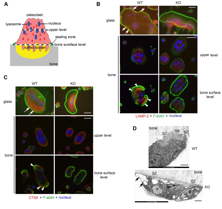
Figure 5. PLEKHM1 regulates lysosome peripheral distribution and ruffled border formation in osteoclasts.
(A) An illustration of the polarized structures of an active osteoclast cultured on bone. The levels (upper level and bone surface level) of two representative sections of confocal microscopic images shown in B and C are indicated. (B) Immunofluorescent staining of LAMP-2 and (C) immunofluorescent staining of cathepsin K (CTSK) in wild-type and Plekhm1–/– (KO) osteoclasts cultured on glass coverslips and bone slices. White arrows in top rows point out the distribution of LAMP-2–positive lysosomes and CTSK at the periphery of WT osteoclasts. White arrowheads in the bottom rows point out the localization of LAMP-2 at the ruffled border and secretion of CTSK in the resorption lacuna inside actin rings of WT osteoclasts cultured on bone. Each image is a representative of 6 coverslips or bone slices/group/culture from at least 3 independent cultures from different mice. Scale bar: 20 μm. (D) Electron microscopic images of WT and KO osteoclasts cultured on bone slices in vitro. Images are representatives of 6 osteoclasts/group. SZ, sealing zone; RB, ruffled border. Black arrows in bottom panel point out enlarged vesicles accumulated in KO osteoclasts. Scale bar: 5 μm.

