
Figure 2. Imaging of FTC/HHS cohort.
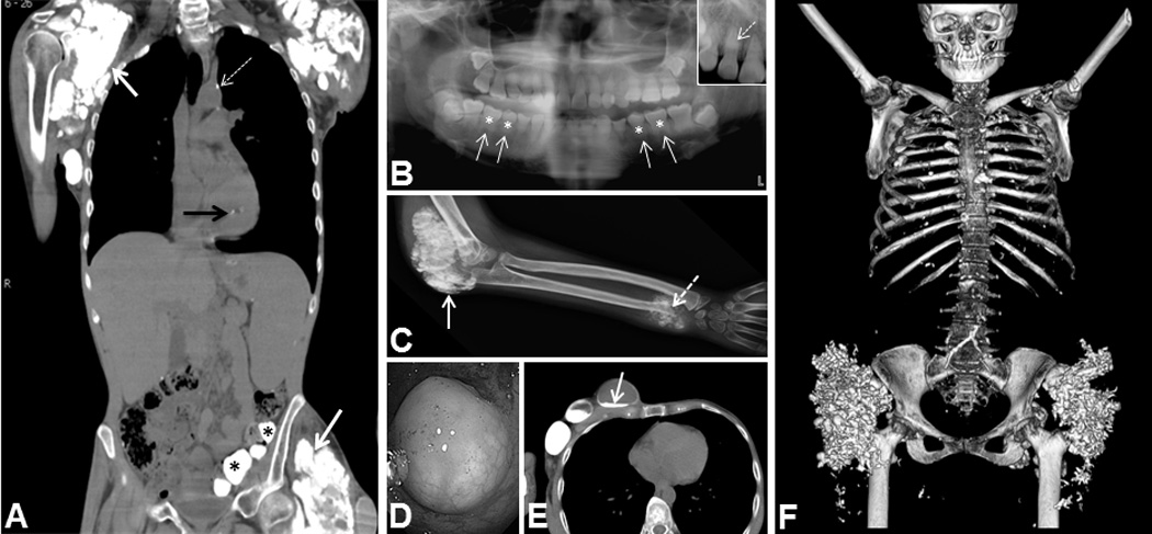
(A) CT showing calcifications in FTC4 including shoulder and hip soft tissue calcifications (white arrows), aortic calcification (dashed arrow), papillary muscle calcification (black arrow), and submucosal gut calcifications (asterisks). (B) Panoramic dental radiograph showing short, bulbous roots (solid arrows) with obliteration of dental pulp (asterisks). Inset - thistle-shaped pulp chambers and a pulp stone (dashed arrow) in FTC5. (C) Radiograph showing tumoral calcinosis of elbow (solid arrow) and ulna with growth plate invasion and destruction in FTC8 (dashed arrow). (D) Submucosal gut calcification on colonoscopy in FTC4. (E) CT showing large cystic masses in the chest wall with fluid levels containing “milk of calcium” in FTC4 (arrow). (F) CT 3-D reconstruction demonstrating extensive calcifications in bilateral hips and scattered calcifications throughout the chest in FTC7.