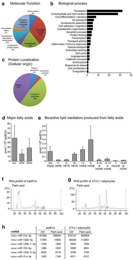
Figure 1. Stressed adipocyte-derived EV protein, lipid, and miRNA composition.
(a–h) 3T3-L1 adipocytes were stressed with 0.5 mM palmitic acid. Stressed adipocyte-derived EVs (sadEVs) were isolated by ultracentrifugation. (a–c) Peptides were resolved by tandem mass spectroscopy and the final protein characterization was performed utilizing UniProt Knowledge Base and BioGPS. Pie charts illustrate (a) molecular function and (c) protein cellular localization; (b) bar graph demonstrates related biological process. See also Supplementary Table 1 for a complete protein composition. (d, e) Characterization of the lipids in the sadEVs by mass spectrometry based lipidomics platform. Total lipids were extracted from purified sadEVs. (d) Bar graph of major fatty acids (in their free, non-esterified formS). AA, arachidonic acid; EPA, eicosapentaenoic acid; and DHA, docosahexaenoic acid. (e) Bar graph of bioactive lipid mediators produced from fatty acids. Dhk-PGD2, dihydro-keto-prostaglandin D2; HEPE, hydroxy-eicosapentaenoic acid; HETE, hydroxy-eicosatetraenoic acid; HODE, hydroxy-octadecadienoic acid; diHOME, dihydroxy-octadecadienoic acid; HDoHE, hydroxy-docosahexaenoic acid; and DiHDPA, dihydroxy-docosapentaenoic acid. (f, g) Characterization of the miRNAs encapsulated in the sadEVs and the corresponding parent 3T3-L1 adipocytes by Agilent 2100 Bioanalyzer. Total RNAs including miRNAs were collected from purified sadEVs or 3T3-L1 adipocytes. (h) Table illustrating differentially expressed miRNAs associated with monocyte/ macrophage activation (raw counts). Palm acid, palmitic acid; Ctrl, control group.

